
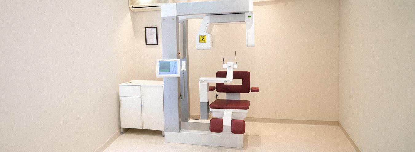
Volumetric Dental CT (Dental Tomography)
Volumetric Dental CT is an advanced radiological imaging method that enables high-resolution, three-dimensional (3D) visualization of the teeth, jawbones, temporomandibular joints, and surrounding anatomical structures. It is an indispensable diagnostic tool, particularly for implant planning and prior to surgical procedures.
What are its features?
- Provides three-dimensional and high-resolution images of the teeth and jaw structures.
- Bone thickness, density, and volume can be assessed through precise measurements.
- Involves a lower radiation dose compared to conventional CT.
- Cross-sectional, panoramic, and 3D reconstructed images can be obtained digitally.
- Patient comfort is high due to the short scanning time.
In which cases is it used?
- Dental implant planning and pre-implant bone assessment
- Analysis of the position of impacted and partially impacted teeth
- Jaw cysts, tumors, and bone lesions
- Assessment of the risk of nerve canal damage
- Temporomandibular joint disorders
- Orthodontic treatment planning
- Examination of the jaw and facial bones following trauma
- Evaluation of root canal anatomy in endodontic problems
How is it performed?
During a Volumetric Dental CT scan, the patient is positioned either standing or seated. The head is stabilized, and the device captures images by making a single rotation around the patient’s head. The scanning time is generally between 20 and 40 seconds. The procedure is painless.


Frequently Asked Questions About Volumetric Dental CT
We have compiled the most frequently asked questions about Volumetric Dental CT (Dental Tomography) for you. Here, you can find answers to questions regarding this advanced imaging method—which provides detailed, three-dimensional visualization of the mouth, teeth, and jaw structures—covering everything from the scanning process and preparation to the indications for use and the evaluation of results.
Volumetrik Diş BT, dişlerin, çene kemiklerinin ve çevre dokuların üç boyutlu görüntülerini elde etmeyi sağlayan gelişmiş bir görüntüleme yöntemidir. Özellikle implant planlaması ve çene yapısının detaylı incelenmesinde kullanılır.
Dental tomografi; implant planlaması, gömülü dişlerin değerlendirilmesi, çene kemiği hastalıkları, ortodontik tedavi planlaması ve çene eklemi problemlerinin incelenmesi gibi birçok durumda kullanılır.
Çekim sırasında hasta cihazın içinde sabit durur ve cihaz başın etrafında kısa bir tur atarak üç boyutlu görüntüler elde eder. İşlem hızlıdır ve genellikle birkaç dakika içinde tamamlanır.
Görüntü alma süresi genellikle 10–30 saniye arasında değişir. Hazırlık süreci ile birlikte işlem birkaç dakika içinde tamamlanır.
Genellikle özel bir hazırlık gerekmez. Ancak çekim sırasında metal takılar, küpe, gözlük veya hareketli diş protezlerinin çıkarılması istenebilir.
Dental tomografi düşük doz X-ışını kullanır ve modern cihazlarda radyasyon miktarı minimize edilmiştir. Bu nedenle gerekli durumlarda güvenle uygulanabilen bir yöntemdir.
Normal diş röntgeni iki boyutlu görüntüler oluştururken, dental tomografi diş ve çene yapısının üç boyutlu görüntülerini sağlar. Bu sayede daha detaylı ve doğru bir değerlendirme yapılabilir.
Görüntüler çekim sonrasında dijital ortamda hemen elde edilir. Radyoloji uzmanı veya diş hekimi tarafından incelenerek kısa süre içinde raporlanır.