
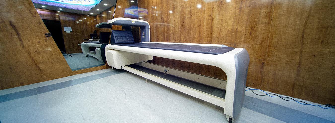
Bone Densitometry (DEXA)
**Bone Densitometry** or **DEXA (Dual Energy X-ray Absorptiometry)** is a specialized imaging method that measures bone mineral density to assess bone strength and fracture risk. It is considered the gold standard for the diagnosis of osteoporosis (bone loss).
What are its features?
- Measures bone mineral density with a high degree of precision.
- Is a reliable and standardized method for diagnosing osteoporosis.
- Uses a very low dose of X-rays.
- Is a fast, painless, and non-invasive procedure.
- Measures can be performed on the spine, hip, and, when necessary, the forearm.
- Allows monitoring of bone loss and treatment progress.
- Provides comparative evaluation through digital measurement and reporting.
- Risk analysis can be performed based on age and sex.
In which cases is it used?
- Diagnosis of osteoporosis and osteopenia
- Assessment of bone density in postmenopausal women
- Routine screening for individuals over 65 years of age
- Patients using corticosteroids or certain hormone medications for an extended period
- Individuals with a family history of osteoporosis or fractures
- Unexplained bone fractures
- Bone monitoring in thyroid, rheumatic, and metabolic diseases
- Monitoring treatment response in patients receiving osteoporosis treatment
- Body fat and muscle mass measurement and monitoring for individuals following a diet and exercise program
How is it performed?
During a Bone Densitometry examination, the patient lies on their back on the examination table. Measurements are generally taken from the lower back and hip areas. The device measures bone density while the patient is asked to remain still. The examination takes approximately 5–10 minutes and is painless.


Frequently Asked Questions About Bone Densitometry (DEXA)
We have compiled the most frequently asked questions about Bone Densitometry (DEXA) for you. Here, you can find answers to questions regarding this method—used to measure bone density and assess the risk of osteoporosis—covering everything from the scanning process and preparation to who should undergo the test and how the results are interpreted.
Kemik Dansitometri, kemik mineral yoğunluğunu ölçmek için kullanılan bir görüntüleme yöntemidir. DEXA (Dual Energy X-ray Absorptiometry) teknolojisi sayesinde kemiklerin ne kadar güçlü olduğu değerlendirilir ve osteoporoz riski belirlenebilir.
DEXA testi genellikle osteoporoz şüphesi olan kişilerde, menopoz sonrası kadınlarda, uzun süre kortizon kullananlarda veya kemik kırığı riski yüksek olan bireylerde yapılır.
Hasta cihazın bulunduğu masaya uzanır ve genellikle bel ve kalça bölgesi taranır. Düşük doz X-ışını kullanılarak kemik yoğunluğu ölçülür ve sonuçlar bilgisayar ortamında analiz edilir.
Kemik dansitometri işlemi genellikle 10–15 dakika içinde tamamlanır. İşlem hızlı ve ağrısızdır.
Çoğu zaman özel bir hazırlık gerekmez. Ancak çekim günü metal aksesuarların çıkarılması istenebilir ve bazı durumlarda kalsiyum takviyelerinin çekimden önce alınmaması önerilebilir.
DEXA testi çok düşük dozda X-ışını kullanır. Bu nedenle güvenli bir yöntem olarak kabul edilir ve birçok hasta için rahatlıkla uygulanabilir.
Test sonucunda kemik yoğunluğu T-skoru ve Z-skoru gibi değerlerle değerlendirilir. Bu değerler kemiklerin normal, düşük yoğunlukta veya osteoporoz seviyesinde olup olmadığını gösterir.
Çekim sonrası elde edilen veriler radyoloji uzmanı tarafından değerlendirilir ve rapor hazırlanır. Sonuçlar genellikle kısa süre içinde doktorunuza iletilir.