
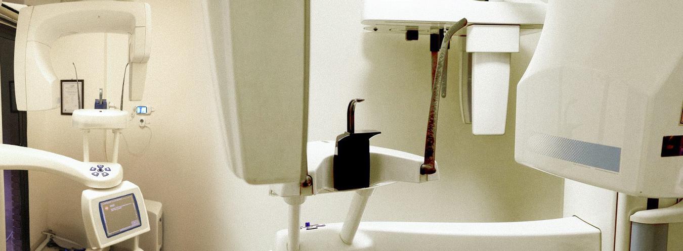
Dental X-Ray (Orthopantomography)
Dental X-Ray (**Orthopantomography**) is a digital radiological imaging method that allows all teeth in the upper and lower jaws, tooth roots, jawbones, and temporomandibular joints to be evaluated in a single image.
What are its features?
- Shows all teeth in the upper and lower jaws in a single image.
- Provides detailed evaluation of tooth roots, jawbones, and temporomandibular joints.
- Clearly determines the position of impacted and partially impacted teeth.
- Provides high-resolution images through digital imaging systems.
- Involves a lower radiation dose compared to conventional X-ray methods.
- Imaging is completed in a short time, allowing results to be obtained quickly.
- Enables digital archiving and comparison with previous images.
In which cases is it used?
- Orthodontic treatment planning
- Pre-implant evaluation
- Impacted wisdom teeth
- Investigation of jaw cysts and tumors
- Tooth root infections
- Jawbone diseases
- Evaluation of the jaw following trauma
- General oral and dental health screening
How is it performed?
During an orthopantomography examination, the patient is positioned standing or sitting inside the imaging device. The head is stabilized, and the device captures images by making a single rotation around the patient’s head. The imaging procedure generally takes 10–15 seconds. The procedure is painless.

Frequently Asked Questions About Dental X-rays
We have compiled the most frequently asked questions about dental X-rays (orthopantomography) for you. Here, you can find answers to questions regarding this imaging method—which plays a vital role in assessing oral and jaw health—covering everything from the procedure and preparation to the indications for use and the interpretation of results.
Ortopantomografi, üst ve alt çene ile tüm dişlerin tek bir görüntüde değerlendirilmesini sağlayan panoramik bir diş röntgeni yöntemidir. Diş hekimleri tarafından tanı ve tedavi planlamasında sıkça kullanılır.
görüntüleme yöntemi; gömülü dişlerin tespiti, çene kemiği hastalıkları, diş kökü enfeksiyonları, ortodontik değerlendirmeler ve implant planlaması gibi birçok durumda kullanılır.
Çekim sırasında hasta cihazın önünde ayakta veya oturarak durur ve çene sabitlenir. Cihaz başın etrafında kısa bir tur atarak tüm çene yapısının panoramik görüntüsünü oluşturur.
Çekim işlemi genellikle birkaç dakika içinde tamamlanır. Asıl görüntü alma süresi ise yalnızca birkaç saniyedir.
Genellikle özel bir hazırlık gerekmez. Ancak çekim sırasında metal takılar, küpe, gözlük veya hareketli protezlerin çıkarılması istenebilir.
Diş röntgeninde kullanılan X-ışını dozu oldukça düşüktür. Bu nedenle tıbbi gereklilik doğrultusunda uygulandığında güvenli kabul edilir.
Ortopantomografi, dişlerin konumu, çene yapısı ve gelişim durumu hakkında geniş bir görüş sağlar. Bu nedenle ortodontik tedavi planlamasında önemli bir rol oynar.
Görüntüler dijital olarak anında elde edilir. Diş hekimi veya radyoloji uzmanı tarafından değerlendirilerek kısa süre içinde sonuçlar paylaşılır.