
What Is Ultrasound? Does It Use Radiation? How Does It Work?
Ultrasound is one of the most commonly used imaging methods in medicine. It is used in many areas, from evaluating abdominal organs to examining the thyroid, breast, soft tissues, and blood vessels. Since it does not involve radiation, can be performed quickly, and provides real-time imaging, it is an important diagnostic tool for both physicians and patients.
What is ultrasound?
Ultrasound, also known medically as ultrasonography, is an imaging method that uses high-frequency sound waves to visualize the internal structures of the body. These sound waves, which cannot be heard by the human ear, are transmitted into the tissues. The reflected waves are detected by the device and converted into images.
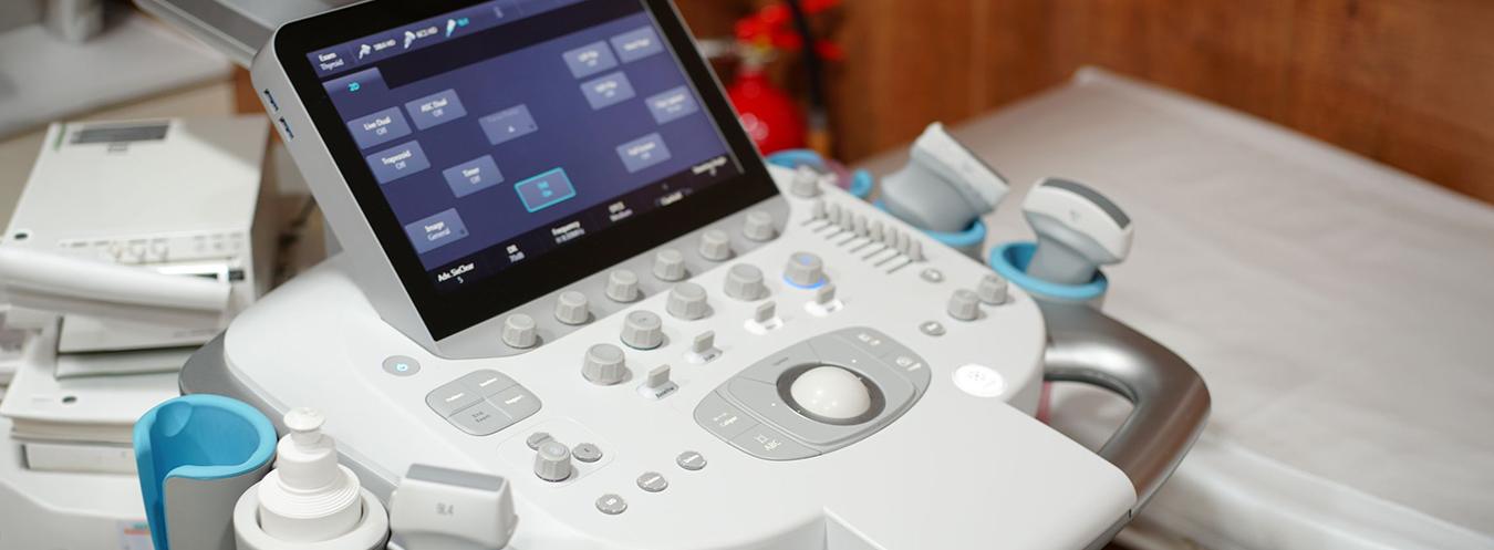
An ultrasound system basically consists of a probe, an image-processing system, and a monitor. The probe is moved over the area being examined, allowing organs, soft tissues, blood vessels, and certain pathological formations to be evaluated in real time.
How does ultrasound work?
The special crystals inside the ultrasound probe convert electrical energy into sound waves. These waves are transmitted into the body and are reflected differently by different types of tissue. The device analyzes the returning signals and creates an image.
Because fluid-filled structures, soft tissues, and dense tissues reflect sound waves differently, organs and lesions can be distinguished from one another. One of the major advantages of ultrasound is that images are obtained in real time.
Is ultrasound harmful?
Ultrasonography does not involve ionizing radiation. Unlike X-rays and computed tomography, it works using sound waves. Therefore, when used appropriately for medical purposes, it is considered safe and is frequently preferred, particularly during pregnancy.
To date, there is no established evidence showing significant harmful effects of diagnostic ultrasound used for medical purposes on human health. For this reason, ultrasound is considered one of the safest imaging methods used in routine medical practice.
Is ultrasound painful or risky?
Ultrasound is generally a painless, quick, and comfortable procedure. Most examinations do not require anesthesia and are usually completed within a short period. Apart from the gel applied to the skin during the examination, there is generally no procedure that causes significant discomfort.
In rare cases, the gel may cause a slight sensation of cold, or minimal discomfort may occur when examining particularly sensitive areas. Apart from this, ultrasound has no known serious side effects.
In which areas is ultrasound used?
Ultrasound has a very wide range of applications. Some of the most common include:
- Examination of the liver, gallbladder, pancreas, and spleen
- Evaluation of the kidneys and urinary tract
- Thyroid and neck ultrasonography
- Breast ultrasonography
- Examination of muscles, tendons, and soft tissues
- Pregnancy monitoring and fetal evaluation
- Examination of blood vessels and blood flow using Doppler ultrasound
Thanks to this wide range of applications, ultrasound provides important information both for diagnosis and follow-up.
How is an ultrasound examination performed?
A special gel is applied to the area being examined. This gel helps transmit sound waves more effectively through the skin. The probe is then moved over the area, and images are obtained.
Some ultrasound examinations may require preparation beforehand. For example, fasting for several hours may be required for an abdominal ultrasound, while some pelvic examinations may require a full bladder. Therefore, it is important to follow the preparation instructions provided before the appointment.
How long does an ultrasound examination take?
Although the duration varies depending on the type of examination, an ultrasound procedure is often completed within 10–20 minutes. It may take slightly longer when a more detailed evaluation is required.
What are the advantages of ultrasound?
The main reasons why ultrasound is widely preferred include:
- Does not involve radiation
- Is painless and fast
- Provides real-time imaging
- Can be safely used during pregnancy
- Allows evaluation of many organs and tissues
Ultrasonography is a safe imaging method that uses high-frequency sound waves, does not involve radiation, and is used to evaluate many medical conditions. Its rapid application, high level of patient comfort, and wide range of applications make it one of the most important diagnostic tools in modern medicine.
What you should know before an ultrasound examination:
- Fasting may be required for some examinations.
- You may be asked to drink water before some pelvic examinations.
- Following the preparation instructions provided before your appointment helps improve image quality.






Frequently Asked Questions About Ultrasonography and Color Doppler
We have compiled the most frequently asked questions about ultrasonography and Color Doppler for you. Here, you can find answers to many questions you might have regarding these safe and rapid imaging methods—covering everything from the scanning process and preparation to the situations in which they are used and the interpretation of results.
Ultrasonografi, yüksek frekanslı ses dalgaları kullanarak vücuttaki organ ve dokuların görüntülenmesini sağlayan bir görüntüleme yöntemidir. Radyasyon içermediği için güvenli kabul edilir ve birçok hastalığın tanısında yaygın olarak kullanılır.
Renkli Doppler ultrasonografi, damar içindeki kan akışını değerlendirmek için kullanılan özel bir ultrason yöntemidir. Kan akışı ekranda renkli olarak gösterilir ve damar tıkanıklıkları veya dolaşım problemleri hakkında önemli bilgiler sağlar.
Ultrasonografi; karın organları, tiroit bezi, meme, kas ve yumuşak dokular gibi birçok bölgenin incelenmesinde kullanılır. Ayrıca gebelik takibi ve bazı girişimsel işlemlerde de önemli bir rol oynar.
Renkli Doppler özellikle damar hastalıkları, varisler, damar tıkanıklıkları, pıhtı oluşumu ve organlara giden kan akışının değerlendirilmesinde kullanılır.
Çekim sırasında incelenecek bölgeye özel bir jel sürülür ve prob adı verilen cihaz cilt üzerinde gezdirilerek görüntüler elde edilir. İşlem genellikle ağrısızdır ve kısa sürede tamamlanır.
İncelenecek bölgeye bağlı olarak bazı durumlarda açlık veya mesanenin dolu olması gerekebilir. Doktorunuz ya da sağlık personeli size gerekli hazırlık hakkında bilgi verir.
Ultrasonografi radyasyon içermediği için güvenli bir görüntüleme yöntemidir. Bu nedenle gebelik döneminde de güvenle kullanılabilmektedir.
Çekim sırasında görüntüler genellikle anında değerlendirilir. Radyoloji uzmanının raporu kısa süre içinde hazırlanarak doktorunuza iletilir.