Computed Tomography (CT) Technology
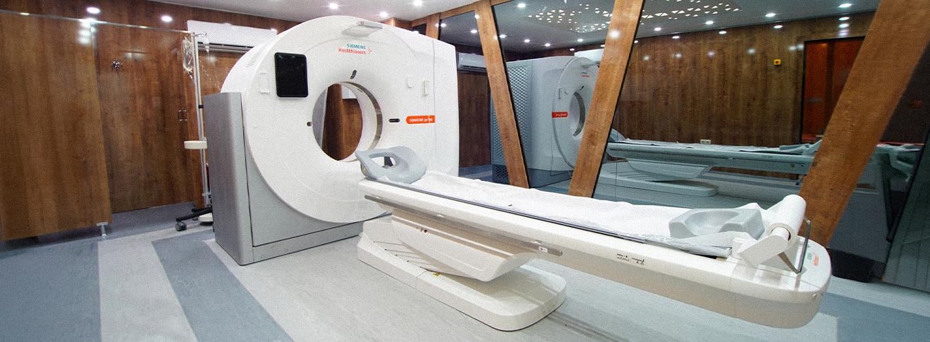
Computed Tomography (CT) is an advanced radiological imaging method that uses X-rays to obtain very thin cross-sectional images of the internal structures of the body. These cross-sectional images are combined using computer technology, allowing detailed evaluation of organs, bones, blood vessels, and soft tissues.
CT plays an important role, particularly in situations where rapid and early diagnosis is required.
What are its features?
- Provides high-resolution and detailed cross-sectional images.
- Imaging can be performed in a very short time.
- Enables rapid diagnosis in emergency situations.
- When necessary, blood vessels and organs can be visualized more clearly using a contrast agent.
- Digital archiving and comparison with previous examinations are possible.
- Low-dose protocols are used with modern CT scanners.
In which cases is it used?
- Cardiovascular diseases (CT angiography)
- Head trauma and suspected brain hemorrhage
- Lung diseases, and evaluation of nodules and embolisms
- Diseases of the liver, pancreas, kidneys, bladder, and peritoneum
- Diseases of the abdominal and pelvic organs
- Investigation of cysts and infections
- Tumor diagnosis and staging
- Kidney stones
- Trauma and multiple injuries
Coronary CT Angiography (Virtual Angiography) is a rapid and detailed CT examination of the blood vessels that supply the heart. Unlike conventional angiography, it does not require access through the groin; the scan is performed using a contrast agent administered through a vein in the arm, and hospitalization is not required. For optimal image quality, heart rate control and compliance with brief breath-holding instructions are important. Patients are generally asked to fast for 4–6 hours before the appointment and avoid caffeine-containing beverages such as tea and coffee for 24 hours. When the contrast agent is administered, a brief sensation of warmth or a metallic taste in the mouth may occur; these effects usually disappear within a short time.

Frequently Asked Questions About Computed Tomography (CT)
Bilgisayarlı Tomografi (BT) hakkında en çok merak edilen soruları sizin için derledik. Çekim öncesi hazırlıklardan işlem sürecine, kontrast maddeden sonuçların alınma süresine kadar aklınızdaki tüm soruların yanıtlarını burada bulabilirsiniz.
CT is an advanced imaging method that uses X-rays to create cross-sectional images of the body. It provides detailed information about tissues, organs, and bone structures.
During the scan, the patient remains still for a short period while images are obtained using X-rays that rotate around the body. The procedure is generally painless and takes only a few minutes.
For some types of CT scans, fasting or drinking a specific amount of fluid may be required. Your doctor will provide you with specific instructions. Metal jewelry should also be removed before the scan.
CT scans use a low dose of X-rays and are generally safe. For pregnant patients or individuals with certain health conditions, your doctor will assess the potential risks and recommend alternative imaging methods if necessary.
Contrast material helps make blood vessels, organs, or lesions appear more clearly. It can be administered orally or intravenously. Its use is generally safe, but caution is required in patients with kidney problems or allergies.
Movement can cause images to become blurry and may make it difficult to make an accurate diagnosis. Therefore, it is very important to remain still throughout the scan.
After the scan, the images are processed by a computer and reviewed by a radiologist. The results are usually provided to your doctor on the same day or within a few days.
While CT uses X-rays, MRI works with magnetic fields and radio waves. CT is faster for imaging bone structures and in emergency situations, whereas MRI is more effective for detailed imaging of soft tissues.