Digital Mammography & Tomosynthesis
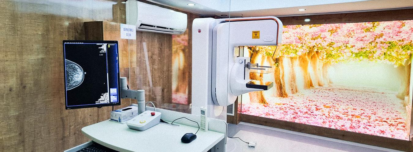
Tomosynthesis Mammography is a new-generation breast imaging method that obtains three-dimensional (3D) images by scanning the breast from different angles using low-dose X-rays. Breast tissue is examined in layers, eliminating uncertainties caused by overlapping structures.
What are its features?
- Advanced tomosynthesis imaging technology
- Excellent resolution and high-speed imaging
- 2D and 3D imaging with artificial intelligence-supported technology
- Imaging system designed to improve patient comfort
- Lower-dose examination options
- Targeted and easier biopsy procedures
- High diagnostic accuracy in dense breast tissue
In which cases is it used?
- Breast cancer screening
- Patients with dense breast tissue
- Palpable breast lumps and breast pain
- Nipple discharge
- Clarification of suspicious mammography findings
- Follow-up after treatment
How is it performed?
The breast is briefly and carefully compressed within the imaging device. Rapid images are acquired from different angles to create three-dimensional images. The procedure is completed within seconds, with patient comfort being a priority.
Breast MRI is an examination that provides a detailed evaluation of breast tissue using a magnetic field and radio waves, without radiation. It is generally requested when mammography or ultrasound is insufficient, for screening women at high risk, or to clarify a finding. In premenopausal women, the appointment is often recommended to be scheduled between days 7 and 14 of the menstrual cycle for optimal image quality. Since an MRI uses a very powerful magnet, metal objects such as jewelry, watches, and credit cards must not be brought into the scanning area. During the examination, the patient lies face down, with the breasts positioned in specially designed openings, and the breasts are not compressed. If necessary, a contrast agent may be administered intravenously.

Frequently Asked Questions About Digital Mammography and Tomosynthesis
We have compiled the most frequently asked questions about digital mammography and tomosynthesis for you. Here, you can find answers to many questions you might have regarding these advanced breast imaging methods—covering everything from the preparation and scanning process to safety and the interpretation of results.
Dijital mamografi, meme dokusunun X-ışınları kullanılarak görüntülenmesini sağlayan modern bir görüntüleme yöntemidir. Meme kanserinin erken teşhisinde en etkili tarama yöntemlerinden biri olarak kabul edilir.
Tomosentez, üç boyutlu mamografi olarak da bilinen gelişmiş bir görüntüleme tekniğidir. Meme dokusunun farklı açılardan çok sayıda görüntüsü alınarak daha detaylı ve katmanlı bir inceleme yapılmasını sağlar.
Dijital mamografi iki boyutlu görüntüler oluştururken, tomosentez meme dokusunun üç boyutlu kesit görüntülerini elde eder. Bu sayede özellikle yoğun meme dokusuna sahip kişilerde daha detaylı değerlendirme yapılabilir.
Mamografi sırasında memenin kısa süreli olarak sıkıştırılması gerekir. Bu durum hafif bir rahatsızlık hissi oluşturabilir ancak işlem genellikle kısa sürer ve tolere edilebilir düzeydedir.
Dijital mamografi çekimi genellikle 10–15 dakika içinde tamamlanır. Tomosentez kullanıldığında işlem süresi biraz daha uzayabilir ancak yine de oldukça kısa sürer.
Çekim günü deodorant, pudra veya krem kullanılmaması önerilir çünkü bu ürünler görüntüleri etkileyebilir. Ayrıca çekim sırasında metal takıların çıkarılması gerekir.
Genellikle 40 yaşından sonra kadınların düzenli olarak mamografi taraması yaptırması önerilir. Risk durumuna göre doktorunuz daha erken yaşta veya farklı aralıklarla tarama önerebilir.
Çekim sonrası görüntüler radyoloji uzmanı tarafından değerlendirilir. Sonuçlar çoğu zaman aynı gün veya kısa süre içinde hazırlanarak doktorunuza iletilir.