
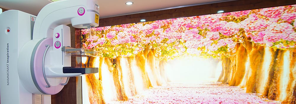
Breast cancer continues to be one of the most common types of cancer affecting women worldwide. In Türkiye, thousands of women receive this diagnosis every year. However, the good news is that when detected at an early stage, breast cancer has a very high treatment success rate. This is where mammography comes into play.
So, when exactly should mammography be performed? At what age should screening begin, how often should it be repeated, and in which cases should additional radiological imaging methods be used?
Although these questions are on the minds of many women, finding clear answers can sometimes be challenging. Different organizations may provide different recommendations, individual risk factors vary from person to person, and continuous advances in radiological technology further add to the complexity. In this comprehensive guide, we will discuss everything you need to know about having mammography performed at the right time and at the appropriate intervals, based on scientific evidence.
What Is Mammography and How Does It Work?
Mammography is a specialized radiological imaging method that uses low-dose X-rays to visualize breast tissue. Its primary purpose is to detect masses that are too small to be felt by hand, calcifications, and other abnormal tissue changes.
During the procedure, the breast tissue is gently compressed between two plates. Although this compression may cause mild discomfort in some women, it generally lasts only a few seconds and is considered safe. Both breasts are imaged from different angles, and the resulting images are evaluated by a radiologist.
Limitations of Mammography
Although mammography is a powerful diagnostic tool, it does not always provide perfect results. Tumors may be obscured, particularly in women with dense breast tissue. This indicates that mammography alone may not always be sufficient and that additional imaging methods may be necessary in some cases.
When Should Mammography Screening Begin?
The appropriate age to begin mammography screening remains a subject of discussion among international medical organizations. However, a general framework can be established.
Recommendations for Women at Average Risk
The Ministry of Health of the Republic of Türkiye and many international organizations recommend the following screening schedule for women at average risk:
- Starting at age 40: Annual mammography screening may be considered. In this age group, the decision to undergo screening is generally based on the woman’s preferences and her physician’s assessment.
- Between ages 45–50: Annual mammography screening is strongly recommended.
- Between ages 50–74: Regular mammography screening should be performed annually or every two years.
- Age 75 and older: The decision to continue screening should be individualized based on the woman’s overall health status and life expectancy.
The American Cancer Society states that women at average risk should undergo annual mammography beginning at age 45 and that, starting at age 55, they may choose to undergo screening every two years or continue with annual screening. On the other hand, some experts and organizations advocate beginning screening at age 40.
Is Mammography Necessary Before Age 40?
Routine mammography is generally not recommended for women under the age of 40. There are two main reasons for this: first, breast tissue tends to be denser in this age group, which can reduce the effectiveness of mammography; second, the incidence of breast cancer is lower among younger women. However, women with certain risk factors may be evaluated differently; this issue will be discussed in greater detail in the following sections.
Mammography Screening Schedule Based on Risk Factors
Not every woman has the same risk of developing breast cancer. Factors such as family history, genetic factors, hormonal status, and lifestyle can significantly affect this risk. Women at high risk may need to begin mammography screening at an earlier age and undergo screening at more frequent intervals.
What Are the High-Risk Factors?
The following conditions may place a woman in the high-risk category for breast cancer:
- Family history: Women with a history of breast or ovarian cancer in a first-degree relative (mother, sister, or daughter)
- Genetic mutations: Individuals carrying BRCA1 or BRCA2 gene mutations
- Personal history of cancer: Women who have previously been diagnosed with breast cancer or lobular carcinoma in situ (LCIS)
- Radiotherapy to the chest: Women who received radiotherapy to the chest between the ages of 10 and 30
- Dense breast tissue: Women whose mammograms show high breast density
- Hormonal factors: Early menarche (first menstrual period), late menopause, and long-term use of hormone replacement therapy
- Pregnancy-related factors: Not having children or having a first birth at a later age
Recommendations for Women at High Risk
Women in the high-risk category are generally advised to begin screening with both mammography and breast MRI from age 30, or even earlier in some cases. For this group, annual mammography combined with MRI is considered the standard approach.
For women at moderate risk, such as those with a family history but without a known genetic mutation, the most appropriate approach is to establish an individualized screening plan with a physician.
Types of Mammography: Digital, 3D, and Beyond
With advances in technology, mammography techniques have undergone significant transformation. Today, there are several options designed to meet different clinical needs rather than a single type of mammography.
Digital Mammography (FFDM)
Digital mammography, which has largely replaced conventional film mammography, records images electronically. This method allows images to be examined more clearly on a computer screen, enlarged, and evaluated using different contrast settings. It has been shown to provide better results, particularly in women under 50 and in individuals with dense breast tissue.
3D Mammography (Tomosynthesis)
Digital breast tomosynthesis, commonly known as 3D mammography, captures breast tissue from multiple angles to create a three-dimensional image. Its main advantages include:
- It can improve the detection of small tumors by allowing individual layers of breast tissue to be examined separately.
- It is more effective in dense breast tissue.
- It reduces the rate of false-positive findings, meaning suspicious findings in the absence of cancer, thereby potentially reducing unnecessary biopsies.
- It increases the breast cancer detection rate.
Although 3D mammography is becoming increasingly widespread, it may not be available at all centers and may involve an additional cost.
Diagnostic Mammography vs. Screening Mammography
There are two main applications of mammography:
- Screening mammography: Mammography performed routinely in women without symptoms for screening purposes. Images are generally obtained using standard views.
- Diagnostic mammography: Mammography performed when a suspicious finding is detected either on physical examination or on a mammogram, providing a more detailed evaluation. Additional views may be obtained, and magnification techniques may be used.
Other Radiological Methods That Complement Mammography
Although mammography is a fundamental method in breast imaging, it may not be sufficient on its own in some cases. In such situations, additional radiological diagnostic tools may be used.
Breast Ultrasonography (USG)
Breast ultrasonography is an important complementary method to mammography, particularly in the following situations:
- Dense breast tissue: Ultrasound can provide additional information in breast tissue that appears dense on mammography.
- Distinguishing cysts from masses: Ultrasound is highly effective in distinguishing fluid-filled cysts from solid masses.
- Young women: It may be preferred during pregnancy and breastfeeding as a radiation-free imaging method.
- Evaluation of palpable masses: It is used to evaluate masses that can be felt by hand but may not be visible on mammography.
Breast MRI
Magnetic resonance imaging (MRI) is one of the most sensitive methods used in breast imaging. However, because of its higher cost, longer examination time, and relatively high false-positive rate in some situations, it is not used as a general screening tool. Breast MRI may be recommended in the following situations:
- Women carrying a BRCA mutation or those with a high genetic risk
- Staging and treatment planning following a breast cancer diagnosis
- Evaluation of implant integrity in women with breast implants
- Indeterminate cases in which mammography and ultrasound do not provide sufficient information
Biopsy: The Path to a Definitive Diagnosis
When a suspicious finding is detected on radiological imaging, a tissue sample is required to establish a definitive diagnosis. Today, biopsies are most commonly performed using minimally invasive, image-guided techniques, such as ultrasound- or mammography-guided biopsy. This allows a definitive diagnosis to be established safely and efficiently without the need for major surgery.
What You Should Know Before and After Mammography
Keeping a few practical considerations in mind can help make the mammography experience more comfortable and efficient.
Preparation Before Mammography
- Consider your menstrual cycle: If possible, schedule your mammogram for approximately one week after the beginning of your menstrual period. During this period, breast tissue may be less tender, making the procedure more comfortable.
- Avoid deodorant and lotions: On the day of the examination, avoid applying deodorant, lotion, cream, or powder to the underarm or breast area. These products may create artifacts on the images.
- Bring previous mammograms: If available, bring your previous mammograms or digital records with you. The radiologist can compare the current images with previous examinations to assess changes more accurately.
- Choose comfortable clothing: Wearing a two-piece outfit can make changing clothes easier.
What Happens After Mammography?
- The radiologist’s evaluation of the results generally takes several days.
- A “recall” does not necessarily indicate a cause for panic; the vast majority of cases requiring additional imaging involve benign findings.
- Be sure to discuss your results with a physician and do not hesitate to ask about anything you do not understand.
Breast Self-Examination: It Does Not Replace Mammography, but It Is Valuable
In addition to regular mammography screening, women are also encouraged to perform monthly breast self-examinations. This can help identify possible changes at an early stage and allow timely consultation with a healthcare professional.
A healthcare professional should be consulted promptly if any of the following findings are noticed:
- A newly developed lump or area of firmness in the breast
- Orange-peel appearance, redness, or swelling of the breast skin
- Nipple retraction, inversion, or discharge
- Swelling in the armpit
- Unexplained breast pain
Timely Screening Can Save Lives
Mammography is a radiological diagnostic method with proven effectiveness in detecting breast cancer at an early stage and can be lifesaving. However, to obtain the greatest benefit from this method, it is important to undergo screening at the appropriate time and at the appropriate intervals.
If you are a woman at average risk, it is generally recommended to begin regular mammography screening from age 40–45 and to establish an individualized screening plan with your physician. If you have high-risk factors, this plan should be developed in your 30s and supported by additional imaging methods when appropriate.
Remember: Mammography is not merely a test; it is a commitment to your own health. Not postponing regular screening, having your results evaluated by a specialist, and using additional imaging methods when necessary are among the most important steps toward early detection.
Do not neglect your health. Consult your physician to determine the next step and schedule your mammography appointment.
This article is intended for general informational purposes only and does not constitute medical advice. Please consult a healthcare professional regarding your individual circumstances and screening schedule.