
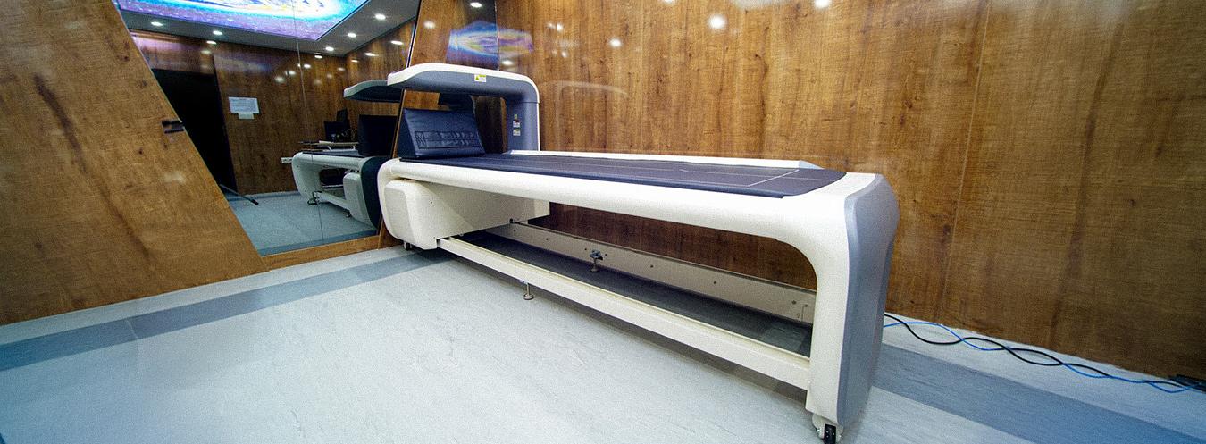
Bone Densitometry (DEXA): What Is It, When Is It Performed, and What Should Be Considered?
Our bones are like the invisible yet indispensable foundation of our body. As we age, our bone structure changes, whether we are aware of it or not. Bone density begins to decrease particularly from our 40s onward, and because this process often progresses silently, it can be difficult to notice. This is where bone densitometry, or DEXA scanning, comes into play. This method is considered a gold-standard diagnostic tool for measuring how strong your bones are in a scientific and reliable manner.
In this article, we will comprehensively discuss what bone densitometry is, when it is necessary, how it is performed, and what should be considered before and after the examination. Having accurate information is one of the most important steps you can take to protect your bone health.
What Is Bone Densitometry (DEXA)?
Bone densitometry, medically known as Dual Energy X-ray Absorptiometry (DEXA or DXA), is a specialized imaging method used to measure bone mineral density (BMD). The term, which can be translated into Turkish as “dual-energy X-ray absorptiometry,” summarizes the working principle of the method: the amount of minerals, particularly calcium, contained in the bones is calculated using two separate X-ray beams at different energy levels.
DEXA scanning is considered the most widely used and reliable method worldwide for diagnosing osteoporosis (bone loss) and osteopenia (bone density below the normal range but not yet reaching the osteoporosis threshold). It is also used to monitor response to treatment and to assess fracture risk.
How Does DEXA Work?
A DEXA device passes two different energy levels of X-rays through the patient’s body. Some of these X-rays are absorbed by bone and soft tissue, while the remainder reaches the other side. The device calculates bone mineral density by comparing the amount of X-rays transmitted and absorbed. The resulting value is expressed in grams per square centimeter (g/cm²).
From a technical perspective, the radiation dose delivered by DEXA is extremely low. Approximately one-tenth of the dose received from a chest X-ray, this amount is considered very low and safe when compared with the natural background radiation we are exposed to in daily life.
Which Areas Are Measured?
The areas most commonly measured with DEXA include:
- Lumbar spine: The L1-L4 vertebrae are particularly evaluated.
- Proximal femur: The total hip and femoral neck can be measured separately.
- Forearm (distal radius): Particularly preferred when hip or spine measurements cannot be performed or when additional information is required.
These areas are among the anatomical sites where fracture risk is highest and where bone loss can be detected at an earlier stage and more clearly.
When Should DEXA Be Performed?
Bone densitometry is not routinely recommended for everyone. Individuals with certain risk factors or those for whom a clinician considers the examination necessary may benefit from this screening.
General Recommendations
The World Health Organization and various national guidelines recommend DEXA screening in the following situations:
- All women aged 65 and older
- All men aged 70 and older
- Women and men aged 50-64 who have certain risk factors
- Postmenopausal women, particularly those who experience early menopause (before age 45)
- Individuals using corticosteroids (cortisone) for an extended period
- Individuals who have previously experienced a low-trauma fracture
- Individuals with inflammatory joint diseases, particularly rheumatoid arthritis
- Individuals with conditions affecting bone metabolism, such as type 1 diabetes, chronic kidney disease, or liver disease
Special Risk Groups
Evaluation for DEXA screening is also recommended for individuals in the following groups:
- Individuals with a low body mass index (BMI < 19 kg/m²)
- Individuals experiencing prolonged immobility (bed rest, paralysis, etc.)
- Individuals with impaired calcium and vitamin D absorption (celiac disease, inflammatory bowel disease)
- Individuals who consume excessive amounts of alcohol and those who smoke
- Individuals with a family history of osteoporosis or hip fracture
- Men diagnosed with hypogonadism (low testosterone)
It is important to note that the indication for DEXA should always be based on an evaluation by a specialist physician. Therefore, even if you believe you fall into one of these categories, consulting your doctor first is the most appropriate approach.
DEXA Examination Methods
Bone densitometry can generally be divided into two main categories: central DEXA and peripheral DEXA.
Central DEXA
Central DEXA is performed using large, table-based devices available in hospitals or imaging centers. It measures clinically critical areas such as the spine and hip. This is the standard method used for the diagnosis of osteoporosis and treatment follow-up.
During the examination, the patient lies on their back on the X-ray table. An imaging arm, known as a collimator, slowly passes over the patient and collects the images. The procedure takes approximately 10-30 minutes and is painless and non-invasive.
Peripheral DEXA (pDEXA)
Peripheral DEXA uses portable, smaller devices to measure areas at the extremities of the body, such as the forearm, wrist, finger, or heel. It is generally used for screening purposes and is preferred primarily to identify individuals at risk rather than to establish a definitive diagnosis. It is less costly than central DEXA and can also be performed in primary healthcare settings.
Quantitative Computed Tomography (QCT)
QCT is an alternative method that measures bone density, particularly in the spine, in three dimensions. Although it involves a higher radiation dose than DEXA, it has the advantage of separately evaluating trabecular (spongy) and cortical (hard outer layer) bone. It is not used as widely as DEXA in routine clinical practice.
How Are DEXA Results Interpreted?
Two key values are included in a DEXA report: the T-score and the Z-score. Understanding these values is essential for correctly interpreting the results.
What Is a T-Score?
The T-score indicates how many standard deviations the measured bone density is above or below the average bone density of a young, healthy adult of the same sex.
According to the definitions of the World Health Organization:
| T-Score | Interpretation |
|---|---|
| -1.0 and above | Normal bone density |
| Between -1.0 and -2.5 | Osteopenia (low bone density) |
| -2.5 and below | Osteoporosis |
| Below -2.5 + presence of a fracture | Severe osteoporosis |
What Is a Z-Score?
The Z-score compares a person’s bone density with that of individuals of the same age, sex, and body structure. The Z-score is considered particularly meaningful in the clinical evaluation of premenopausal women, men, and children. When the Z-score is below -2.0, bone loss beyond what would be expected for the person’s age may be present, and underlying causes should be investigated.
What Should Be Considered Before and After a DEXA Examination?
Preparation is important for obtaining an accurate and reliable measurement. Here are the key points to consider:
What Should Be Considered Before the Examination?
Calcium supplements and medications: Stop taking calcium supplements at least 24 hours before your appointment. Calcium may accumulate on the surface of the bones and artificially increase the measurement. However, do not stop any other medications prescribed by your doctor without consulting your doctor.
Metal accessories and clothing: You may be asked to remove belts, zippers, piercings, and jewelry containing metal during the examination. Therefore, wearing comfortable clothing that can be easily removed on the day of the examination can make the process easier.
Contrast agents or radioactive substance injections: If you have undergone a contrast-enhanced CT scan, scintigraphy, or barium examination within the previous 7-10 days, residual contrast material may affect the DEXA result. Inform the relevant department about this before your appointment.
Pregnancy: Although DEXA involves a low dose of radiation, it is not routinely recommended during pregnancy. Be sure to inform the medical team before the examination if you are pregnant or think you may be pregnant.
Fasting is not required: No special fasting period is required for DEXA; you can maintain your normal eating routine.
What Should Be Considered During the Examination?
- Follow the technician’s instructions and move slowly when asked to change position.
- Your legs may be stabilized with a special support device; this is completely normal.
- The technician will let you know if you need to hold your breath.
- If you experience pain or discomfort, ask for the procedure to be stopped and inform the technician.
What Should Be Considered After the Examination?
Since DEXA is a non-invasive procedure, it does not require a special recovery period. You can immediately return to your normal daily activities after the examination. Rather than attempting to interpret your results on your own, it is much more appropriate to evaluate them together with the physician who is following your care.
Frequently Asked Questions About DEXA
How often should DEXA be performed?
This depends entirely on the clinical situation and risk factors. In general, repeat examinations may be recommended every 1-2 years for patients receiving treatment, while intervals of 2-5 years may be sufficient for low-risk individuals.
Can cancer be diagnosed with DEXA?
No. DEXA measures only bone density. However, some devices can also perform body composition analysis, such as measuring fat mass and muscle mass.
Is DEXA harmful?
The radiation dose from DEXA is extremely low. Even the cosmic radiation exposure during a flight between Istanbul and Ankara can be comparable to the dose received from a DEXA scan. Therefore, it is considered safe for routine use.
Is special preparation required?
No additional preparation is required when the precautions mentioned above are followed. Adequate sleep and a normal diet are sufficient.
Conclusion: Take Your Bone Health Seriously
Although osteoporosis is often regarded as an inevitable part of aging, it can in fact be slowed and largely controlled with appropriate treatment and preventive measures. The first step is accurate diagnosis. DEXA scanning is one of the most sensitive, safe, and accessible methods currently available for measuring bone density.
If you have risk factors or your doctor considers the examination necessary, do not postpone the scan. Remember that measures taken after a fracture do not produce the same results as measures taken before a fracture occurs. Early diagnosis does not simply provide you with a report consisting of a number; it offers a realistic and personalized roadmap for protecting your bone health.
Take good care of your bones. Because everything that keeps you standing depends on them.
This article has been prepared for informational purposes only. Please consult a specialist physician for any diagnosis or treatment decision.