
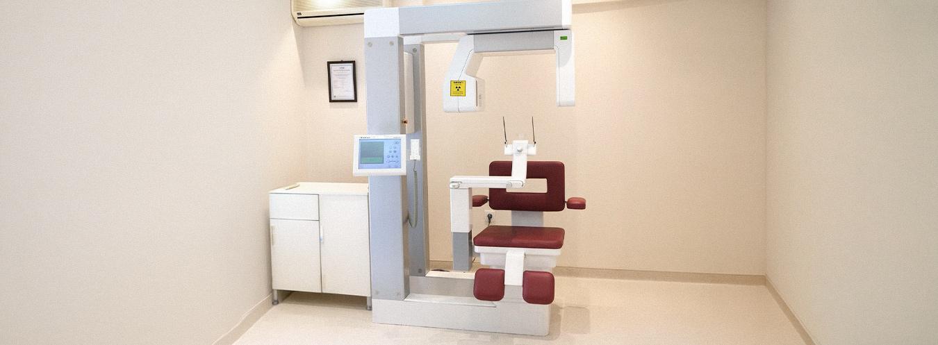
Volumetric Dental CT (Dental Tomography)
Volumetric Dental CT is an advanced radiological imaging method that enables high-resolution, three-dimensional (3D) visualization of the teeth, jawbones, temporomandibular joints, and surrounding anatomical structures. It is an indispensable diagnostic tool, particularly for implant planning and prior to surgical procedures.
What are its features?
- Provides three-dimensional and high-resolution images of the teeth and jaw structures.
- Bone thickness, density, and volume can be assessed through precise measurements.
- Involves a lower radiation dose compared to conventional CT.
- Cross-sectional, panoramic, and 3D reconstructed images can be obtained digitally.
- Patient comfort is high due to the short scanning time.
In which cases is it used?
- Dental implant planning and pre-implant bone assessment
- Analysis of the position of impacted and partially impacted teeth
- Jaw cysts, tumors, and bone lesions
- Assessment of the risk of nerve canal damage
- Temporomandibular joint disorders
- Orthodontic treatment planning
- Examination of the jaw and facial bones following trauma
- Evaluation of root canal anatomy in endodontic problems
How is it performed?
During a Volumetric Dental CT scan, the patient is positioned either standing or seated. The head is stabilized, and the device captures images by making a single rotation around the patient’s head. The scanning time is generally between 20 and 40 seconds. The procedure is painless.


Frequently Asked Questions About Volumetric Dental CT
We have compiled the most frequently asked questions about Volumetric Dental CT (Dental Tomography) for you. Here, you can find answers to questions regarding this advanced imaging method—which provides detailed, three-dimensional visualization of the mouth, teeth, and jaw structures—covering everything from the scanning process and preparation to the indications for use and the evaluation of results.
Cone Beam Computed Tomography (CBCT), also known as Dental CT, is an advanced imaging method that provides three-dimensional images of the teeth, jawbones, and surrounding tissues. It is particularly used for implant planning and detailed examination of the jaw structure.
Dental CT is used in many situations, including implant planning, evaluation of impacted teeth, jawbone diseases, orthodontic treatment planning, and the examination of temporomandibular joint (TMJ) problems.
During the scan, the patient remains still inside the device while it makes a short rotation around the head to obtain three-dimensional images. The procedure is quick and is usually completed within a few minutes.
The image acquisition time usually ranges from 10–30 seconds. Including the preparation process, the examination is completed within a few minutes.
Generally, no special preparation is required. However, you may be asked to remove metal jewelry, earrings, glasses, or removable dental prostheses during the scan.
Dental CT uses a low dose of X-rays, and modern devices are designed to minimize radiation exposure. Therefore, it is a method that can be safely performed when medically necessary.
While a conventional dental X-ray produces two-dimensional images, dental CT provides three-dimensional images of the teeth and jaw structures. This allows for a more detailed and accurate evaluation.
The images are obtained digitally immediately after the scan. They are reviewed by a radiologist or dentist and reported within a short period.