
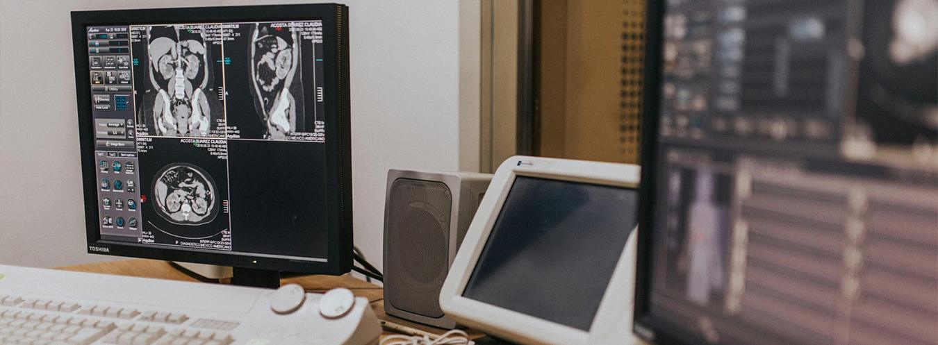
Magnetic Resonance (MRI) Imaging
Magnetic Resonance (MRI) is an advanced imaging method that uses powerful magnetic fields and radio waves to obtain detailed images of the body. MRI does not use X-rays or ionizing radiation, making it a safe option for many patient groups. It provides high diagnostic accuracy, particularly in the evaluation of soft tissues.
What are its features?
- Does not involve ionizing radiation.
- Provides superior soft tissue imaging.
- Provides multiplanar images.
- Provides detailed imaging of the brain, spinal cord, muscles, ligaments, and joints.
- When necessary, a more detailed examination can be performed using a contrast agent.
- Provides digital archiving and the opportunity for comparative follow-up.
- Modern MRI systems are designed to improve patient comfort.
In which cases is it used?
- Brain and spinal cord diseases
- Herniated discs in the lower back and neck
- Tumor and mass characterization and local staging
- Neurological diseases (MS, epilepsy, etc.)
- Liver, biliary tract, and pancreatic diseases
- Small bowel diseases and defecation disorders
- Kidney, prostate, and gynecological conditions
- Joint, ligament, and meniscal injuries
- Evaluation of heart and vascular diseases
Cardiac MRI; is an MRI examination that provides a detailed evaluation of the structure and function of the heart without the use of ionizing radiation. During the examination, ECG electrodes are attached to the chest, and images are synchronized with the heartbeat. Short breath-holding instructions are given at frequent intervals; remaining still is important for image quality. In some protocols, a contrast agent and/or stress medications may be administered intravenously. Since MRI uses a very powerful magnet, information about metal implants and cardiac devices such as pacemakers or ICDs is critical for safety.


Frequently Asked Questions About Magnetic Resonance (MRI)
We have compiled the most frequently asked questions about Magnetic Resonance (MR) imaging for you. Here, you can find answers to many questions you might have—ranging from pre-scan preparations and what happens during the procedure to safety considerations and the interpretation of results.
Magnetic Resonance Imaging (MRI) is an advanced imaging method that uses a powerful magnetic field and radio waves to create detailed images of the internal structures of the body. It is commonly used to examine the brain, spine, muscles, joints, and soft tissues.
During an MRI scan, the patient lies on a movable table that slides into the MRI machine and is asked to remain still throughout the scan. The machine uses a magnetic field and radio waves to create cross-sectional images. The procedure is generally painless.
Depending on the area being examined, an MRI scan usually takes between 15 and 45 minutes. In some detailed examinations, the procedure may take a little longer.
During an MRI scan, rhythmic and loud noises may occur due to changes in the magnetic field as the machine operates. These sounds are normal, and patients are usually provided with headphones or earplugs to reduce discomfort.
Most MRI scans do not require any special preparation. However, metal jewelry, watches, credit cards, and other items that may be affected by the magnetic field must be removed. In some cases, your doctor may ask you to fast before the scan.
Since MRI does not use X-rays, it is generally considered a safe imaging method. However, patients with pacemakers, metal implants, or certain prostheses must inform the medical staff before the scan.
In some MRI examinations, contrast material may be used to provide clearer images of blood vessels, tumors, or inflamed tissues. This material is usually administered intravenously and allows the images to be evaluated in greater detail.
MRI images are reviewed and reported by a radiologist after the scan. The results are usually provided to your doctor on the same day or within a few days.